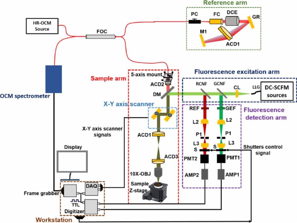
WOBURN, MA, JANUARY 24, 2022 — Biology researchers at Indiana University1 have developed an integrated system combining high-resolution optical coherence microscopy (HR-OCM) with dual-channel scanning confocal fluorescence microscopy (DC-SCFM) to enable 3D visual evaluation of cell activities involved in pupil developmental and disease conditions. Still in its experimental stages, this dual-modality 3D system simultaneously co-registers reflectance and fluorescence signals, giving it the ability to accurately track structural and functional changes in live specimens over time. Indiana University researchers hope to use their system to enable new investigations of biological processes in small animal models.
A BitFlow Axion Camera Link frame grabber is a critical component of the hybrid system. It acquires the output signal from a spectrometer equipped with a Teledyne e2v high-speed line-scan camera operating at the rate of 250 kHz. A lateral resolution of 2-μm and axial resolution of 2.4-μm is captured in tissue over a field-of-view of 1.1 mm ×1.1 mm. The analog scanning signals, as well as the trigger signals for the BitFlow frame grabber, are generated synchronously through a four-channel analog output data acquisition card. Simultaneous recording of HR-OCM and DC-SCFM data was performed using custom software developed in LabVIEW 2017.
As data generated by faster, higher-resolution Camera Link cameras continues to grow exponentially, the Axion’s PCIe Gen 2 interface, with its StreamSync™ DMA optimized for modern computers, is needed to optimize their full performance. Features such as easier switching between different tap formats, a powerful acquisition engine, and a more flexible I/O and timing generator are all readily available in a dedicated low cost CL Base orientated frame grabber.
During development, researchers applied different strategies to enable the simultaneous recording of information, as well as to overcome the focal plane mismatch between both imaging modalities. The system’s performances were evaluated in imaging fluorescence microspheres embedded in multi-layer tape and silicone phantom.
The combined system is synergistic in generating structural and functional information of samples; the DC-SCFM allows for the discrimination between different fluorophores, while the HR-OCM enables the 3D localization of the features inside tissue samples and enabled the depth localization.
1 “Development of high-speed, integrated high-resolution optical coherence microscopy and dual-channel fluorescence microscopy for the simultaneous co-registration of reflectance and fluorescence signals” Reddikumar Maddipatla, PatriceTankam School of Optometry, Indiana University, Bloomington, IN 47405, USA